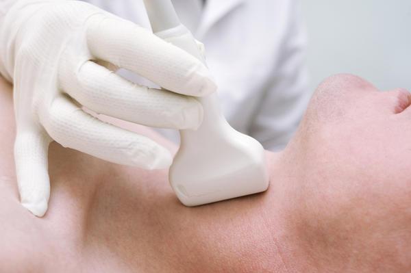
Thyroid scintigraphy
Under scintigraphy we mean the method of functionaldiagnostics, which consists in the introduction into the body of substances possessing radioactive properties. The result is an image, by determining the level of radiation emitted.
Scanning of thyroid tissue is intended to determine the functional activity of its cells by the amount of capture of substances with radioactive properties.
In the course of its functioning it absorbsiodine, other substances necessary for the production of hormones. The speed of this process is not constant. At different times it can increase or decrease. Therefore, the magnitude of consumption of these substances is an indicator of the functional state of the thyroid gland.
Assessment of the functional state is carried out atassistance in the introduction into the body of special substances with radiological activity, which are then distributed throughout the tissues of the body. The thyroid gland absorbs a sufficiently large number of such elements, yielding only sexual and salivary glands in this indicator.
In order to understand what scintigraphy isthyroid gland, it is necessary to present this organ as a filter for substances. They pass through it and in this form settle in the bloodstream. This dependence is observed: with a good filter functionality, all substances, including radioisotopes, are better retained in it. With its passivity, the opposite phenomenon is observed (all elements falling into it are not filtered, but fall directly into the blood).
Thyroid scintigraphy isfunctional diagnostic method aimed at determining the degree of saturation of its tissues with radioactive isotopes. This indicator allows you to judge the functional activity of the organ.
Scanning helps to identify indirectlyfeatures of the structure. The contours of the thyroid gland determines its size, its relation to the most closely located structures in the neck. Also, the degree of absorption of radioactive substances determines the state of the focal formation. But such a study as thyroid scintigraphy can not be called exact, it is indicative. This is because the basis of scintigraphy is the determination of the functional state of the gland.
This study, as well as kidney scintigraphy,requires the use of the following radioisotopes: iodine-123, technetium-99t pertechnetate. They are administered intravenously, in rare cases through the mouth. After their introduction, the patient moves to the gamma camera, which receives radiation from the tissues of the thyroid gland. It is also called a medical scanner. Scintigraphy of the thyroid gland ends with the results, which are fixed on colored paper.
The image reflects the lower part of the head with the neck, as well as the upper part of the chest.
The background density indicates a sufficiently hightissue saturation with radioisotope substances. With this result, a conclusion is made about high functional activity. In the presence of a light background, they speak of a decrease in the function of the gland or its site. In medical practice, the terms "cold" and "hot" are used to indicate the density of foci. These symbols are characteristic for all types of research (among them, liver scintigraphy). Under normal functioning, the organ should be uniformly saturated with a radioisotope, its areas should not have focal blackouts.
Thus, scintigraphy of the thyroid glandis an important diagnostic method that allows you to assess the functional state of this organ and determine such pathological conditions as hypothyroidism, thyrotoxicosis.
</ p>